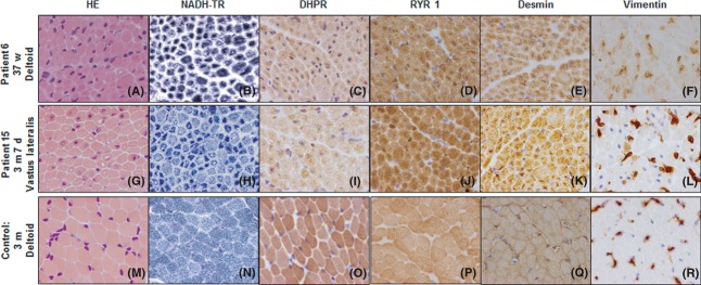
Figure 1.

Transverse muscle sections of Patients 6 and 15 with severe X-linked myotubular myopathy showing marked variability in fiber size and the presence of numerous hypotrophic myofibers with centrally placed nuclei (haematoxylin-eosin [HE]; pictures A and G). The central area of some muscle fibers showed increased oxidative enzyme activity staining; a pale halo in subsarcolemmal regions is also observed (NADH-tetrazolium reductase [NADH-TR]; pictures B and H). Muscle sections demonstrate fibers with positive expression of either DHPR (Pictures C and I), RYR1 (Pictures D and J), or desmin (Pictures E and K) with a labeling increased in the central areas of the fibers. The immunolabeling of Vimentin is observed in some fibers (Pictures F and L). Control muscle sections (Pictures M–R). d, days; w, weeks; m, months; DHPR, dihydropyridine receptor-a1subunit; RYR, ryanodine receptor type 1.
