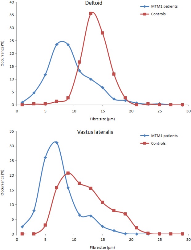
Figure 3.

Meta-distribution of muscle fiber size observed in X-linked myotubular myopathy (XLMTM) patients and controls for the deltoid and the vastus lateralis muscles. The histograms were normalized to unit area; the amplitude in each bin is thus expressed as a percentage of occurrences. The graph shows an obvious shift in myofiber size distribution toward smaller diameters, in both vastus lateralis and deltoid, in all patients.
