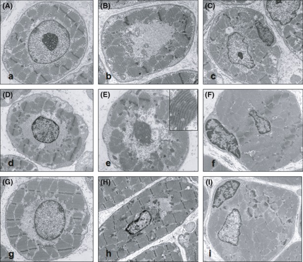
Figure 4.

Ultrastructural analysis of muscle biopsies from Patient 4 (A and B), Patient 5 (C), Patient 7 (D and E), Patient 8 (F), Patient 12 (H), and Patient 13 (G and I). The transverse sections show myonuclei in the center of the fiber often bordered by mitochondria, glycogen, and tubular structures (Pictures A, C, D, F–I). In sections crossing the muscle fibers between two adjacent nuclei, the central area of the fibers displays a reduction of myofilaments; the space is occupied by mitochondria, glycogen, and tubular structures (Pictures B, E). Numerous cisterns corresponding to endoplasmic reticulum, triads, and Golgi complexes are found especially in the central areas of the fibers (Pictures B, E, H). Longitudinal sections for visualizing the mitochondria and glycogen that aggregate around the poles of the nucleus (E). The satellite cells are located beneath the basal lamina that surrounds each fiber. The nucleus of the satellite cells usually appears darker (with condensed chromatin) than the central nucleus in the muscle fibers (Pictures C, F, I).
