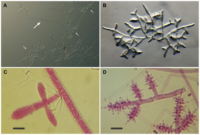
Figure 2. Escovopsis moelleri.
(A–B) Growth habit, note stolons (long arrow) formed at the colony edge with rhizoids (short arrows) developing on the agar surface; (C–D) Details of conidiogenesis showing early development of the clavate vesicles (C, scale bar = 10 µm), and, a later stage covered with swollen, short-necked phialides (D, scale bar = 20 µm).

