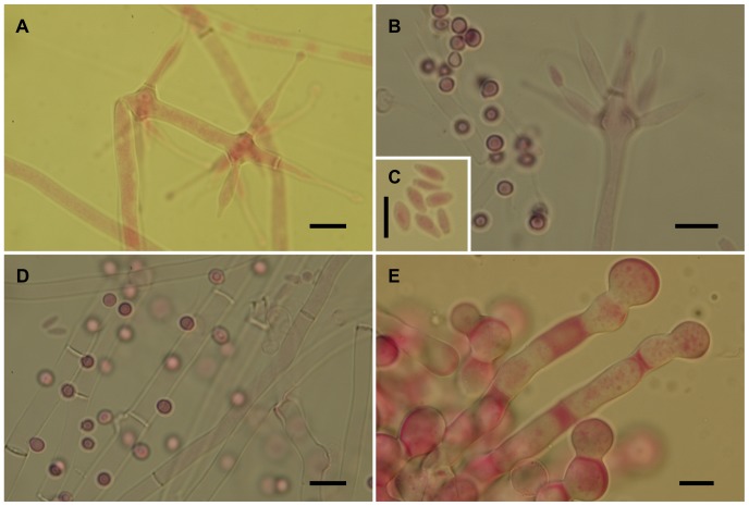
Figure 6. Escovopsioides nivea.
(A–B) Conidiophores bearing both terminal and intercalary vesicles with few cylindrical, subulate phialides tapering gradually to a long neck region, and hyaline, thin-walled conidia (inset, C)—distinguished from the sphaerical darker aleurioconidia (B, left above inset); (D) Aleurioconidia emerging directly from hyphae; (E) Chlamydospores sensu lato formed in glistening white chains or ropes, densely guttulate. All scale bars = 10 µm.

