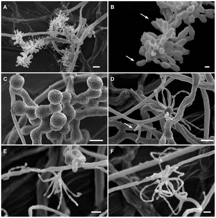
Figure 7. Details of conidiogenesis and spore morphology, as revealed by Critical-Point Drying SEM.
(A) Escovopsis moelleri, showing branching and vesicle formation (scale bar = 10 µm); (B) Detail of conidial morphology, with ornamentation and apical cap (arrows) (scale bar = 2 µm); (C) Escovopsioides nivea, chains of chlamydospores sensu lato revealing cryptic surface ornamentation or mucilaginous deposit (scale bar = 10 µm); (D–F) Escovopsioides nivea, (D) showing both terminal vesicle and phialides produced laterally on slight swelling (arrow) (scale bar = 10 µm); (E–F) Lageniform phialides with long chains of conidia (scale bar = 5 µm).

