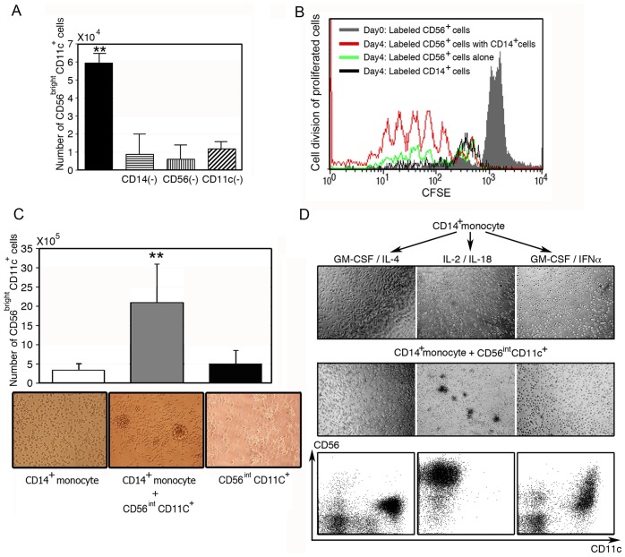
Figure 3. Role of CD14+ monocytes in the development of CD56brightCD11c+ cells.
(A) CD14+, CD56+, and CD11c+ cells were required for the development of CD56brightCD11c+ cells. CD14+, CD56+, or CD11c+ cells were depleted from T cell-depleted PBMCs (2×10⁴ cells/0.5 ml) and stimulated with IL-2/ IL-18 for 7 days. The number of CD56brightCD11c+ cells was counted based on trypan blue dye exclusion. Data show mean ± SD (n = 5), **p<0.01. (B) Effect of CD14+ monocytes on the division of CD56+ cells. CFSE-labeled CD56+ cells were cultured with purified CD14+ monocytes at a ratio of 1:1 at a cell density of 2×10⁵/well. After co-culture for 4 days, proliferating cells were analyzed by CFSE, CD56 and CD11c expression. A representative result of three independent experiments is shown. Division of CFSE-labeled CD14+ cells is also shown (black line). (C) Formation of large aggregates of CD56brightCD11c+ cells in the co-culture of CD56intCD11c+ cells and CD14+ monocytes in the presence of IL-2/IL-18 for 7 days, not in cultures of individual cell types. Data show mean ± SD (n = 4), **p<0.01, and the morphological data are representative of three independent experiments. (D) IL-2/IL-18-dependent cell aggregation in culture of CD14+ monocytes and CD56intCD11c+ cells. IL-4-DCs, IL-18-induced CD14+ monocytes and IFN-α-DCs were co-cultured with freshly isolated CD56intCD11c+ cells (1×10⁵ cells/well) at a ratio of 1:1, respectively. After 12 h incubation, cell aggregates were observed under a microscope. Flow cytometric analyses are carried out after 3 days of co-culture. A representative result of three independent experiments is shown.

