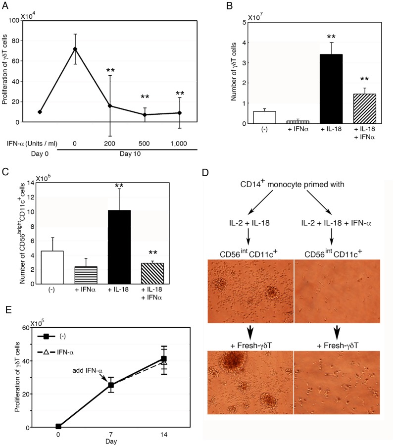
Figure 5. Negative regulation of CD56brightCD11c+ cell development by IFN-α.
(A) Inhibition of γδ T cell proliferation by IFN-α in PBMC cultures. PBMCs were stimulated with ZOL/IL-2 for 10 days in presence of various doses of IFN-α. Data show mean ± SD (n = 5), **p<0.01. (B) Attenuation by IFN-α of IL-18-mediated γδ T cell expansion in PBMC cultures with ZOL/IL-2. Data show mean ± SD (n = 4), **p<0.01. (C) Abrogation by IFN-α of the development and proliferation of CD56brightCD11c+ cells in cultures of CD3+ T cells-depleted PBMCs. Data show mean ± SD (n = 4), **p<0.01. (D) Inhibition of cell aggregation by IFN-α in cultures of CD56intCD11c+ cells and CD14+ monocytes, in the absence and presence of freshly isolated γδ T cells. CD14+ monocytes were pretreated with IL-2/IL-18 for 3 days, with or without IFN-α then CD56intCD11c+ cells were added into the culture (upper panels). Next, freshly isolated γδ T cells were added and cellular clusters were observed by microscope (lower panels). The cell aggregates image is representative of three independent experiments. (E) Proliferation of γδ T cells in cultures containing mature CD56brightCD11c+ cells even in the presence of IFN-α. Freshly isolated γδ T cells and mature CD56brightCD11c+ cells were stimulated with ZOL/IL-2, with or without further addition of IFN-α since day 7 onwards and were continuously incubated. The number of proliferating cells was assayed after another 7 days' culture. Data show mean ± SD (n = 5).

