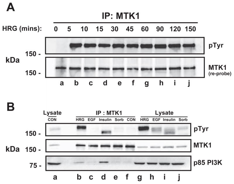
Fig. 3.
Sustained MTK1 and HER3 association. MCF-7 cells were stimulated with 10 nM HRG for the indicated times. MTK1 was immunoprecipitated and proteins were resolved by SDS-PAGE, then immunoblotted with anti-phosphotyrosine antibody (A, top panel). The membrane was re-robed with MTK1 antibody (bottom panel). T-47D cells were stimulated with 10 nM HRG for 12 min, 3.3 nM EGF for 12 min, 10 μg/ml insulin for 15 min or 0.3 M sorbitol for 30 min followed by immunoprecipitation of MTK1. The proteins were resolved by SDS-PAGE and immunoblotted with the indicated antibodies.

