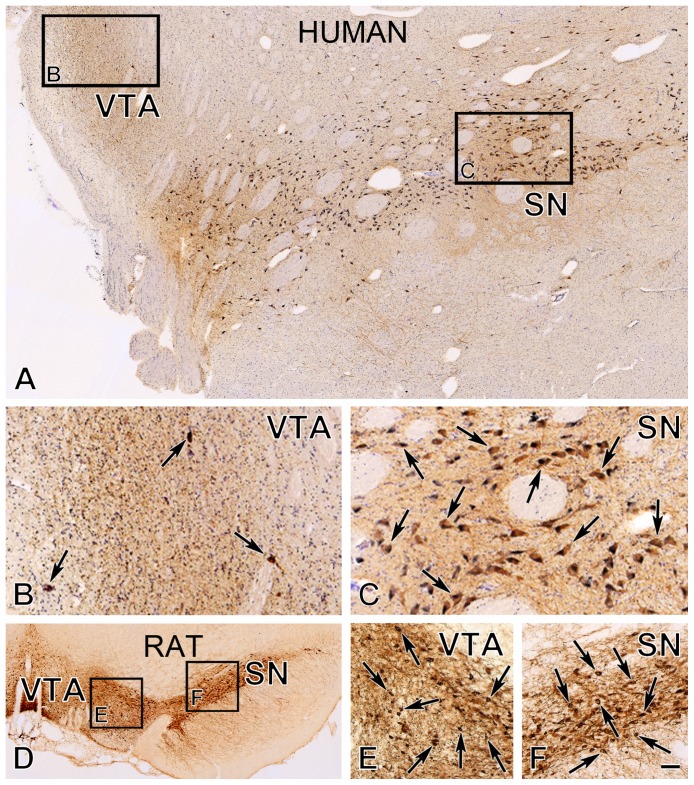
Figure 1. Immunohistochemical detection of dopaminergic neurons in the VTA and the SN of the human and the rat.
Representative low-power images of immunostained sections from an adult male human (A) and rat (D) illustrate the distribution of tyrosine hydroxylase (TH)-immunoreactive (IR) dopaminergic neurons in the ventral tegmental area (VTA) and pars compacta of the substantia nigra (SN). Medium-power images (insets B, C, E and F) reveal that the pars compacta of the SN contains densely-packed dopaminergic neurons (arrows) in both species. In contrast, while dopaminergic neurons are distributed loosely in the human VTA (arrows in B), they exhibit a relatively high regional cell density in the VTA of the rat (arrows in E). Cresyl violet staining in A–C visualizes non-dopaminergic perikarya. Scale bar = 200 µm in A, D and 66 µm in B, C, E, F.

