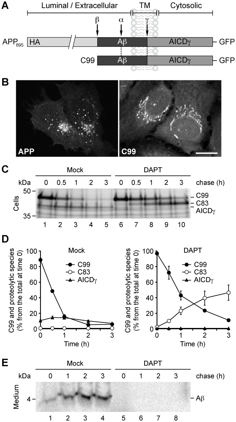
Figure 1. Intracellular localization and proteolytic processing of C99.
(A) Schematic representation of GFP-tagged APP and C99 indicating their topological domains and the position of the HA tag, the Aβ peptide, the proteolytic cleavage sites (α, β and γ), and the AICDγ fragment. (B) Fluorescence microscopy analysis of H4 human neuroglioma cells transiently expressing APP-GFP or C99-GFP. Bar, 10 µm. (C–E) H4 cells transiently expressing C99-GFP were left untreated or treated for 16 h with 1 µM DAPT, labeled for 4 hr at 20°C with 1 mCi/ml [35S]-methionine-cysteine, and chased at 37°C for the indicated times. C99 and Aβ species were immunoprecipitated from cell lysates with anti-GFP antibody (C), or from the culture medium with 6E10 antibody (E), respectively. Proteins were analyzed on 10%–20% Tricine gels and fluorography. The positions of molecular mass markers are indicated on the left. (D) Densitometric quantification of the levels of C99, C83, and AICDγ shown in C.

