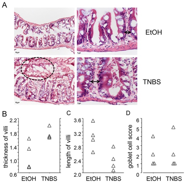
Figure 3. Histological features of TNBS-induced enterocolitis in adult zebrafish.

A) Representative H&E stained sections form TNBS- or vehicle (30% ethanol) treated animals, at two different magnifications. Arrowheads indicate villi thickness in TNBS- and vehicle-treated intestinal sections. A region showing luminal sloughing of cellular debris in the TNBS-treated intestine is marked by a dotted line.
B-C). Relative villi thickness and height (in mm) were evaluated as described in Methods.
D) Abundance of goblet cells based on a 0-5 scoring system.
