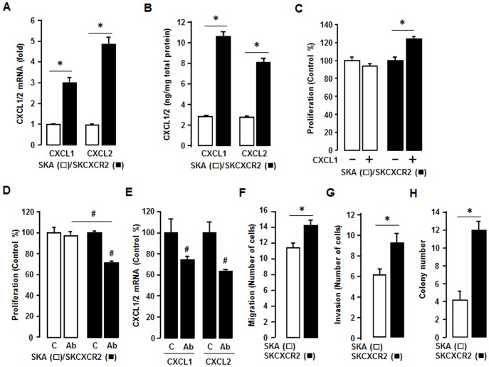
Figure 3. SKCXCR2 cells increases CXCL1/2, thus contributing to cell proliferation and enhancing migration, invasion and colony formation relative to values in SKA cells.
(A) Confirmation of CXCL1 and CXCL2 expression in SKA and SKCXCR2 cells by qRT-PCR. After isolating total RNA, qRT-PCR was carried out using primers for CXCL1 and CXCL2. (B) Cellular CXCL1 and CXCL2 concentrations in SKA and SKCXCR2 cells over a period of 24 h. Whole cell lysates were prepared and ELISA carried out using antibodies specific to CXCL1 and CXCL2 and values were normalized to total protein. (C) Effect of CXCL1 on cell proliferation in SKA and SKCXCR2 cells for 48 h incubation. (D) Effect of pan antibody for CXCL1/2/3 on cell proliferation in SKA and SKCXCR2 cells. Cells were incubated with normal IgG (Control) and pan antibody (1∶100 dilution) for 48 h. The cell proliferation assay was performed using MTT and values were normalized to untreated controls. (E) Effect of pan antibody for CXCL1/2/3 on CXCL1 and CXCL2 expression in SKCXCR2 cells by qRT-PCR. After treating with pan antibody for 24 h and then isolating total RNA, qRT-PCR was carried out using primers for CXCL1 and CXCL2. (F) Migration characteristics between SKA and SKCXCR2 cells. (G) Invasion characteristics between SKA and SKCXCR2 cells. (H) Comparison of colony formation between SKA and SKCXCR2 cells. All experiments were performed at least in triplicate and data are shown as mean ± S.E. * and # (p≤0.05) as calculated by Student’s t-test.

