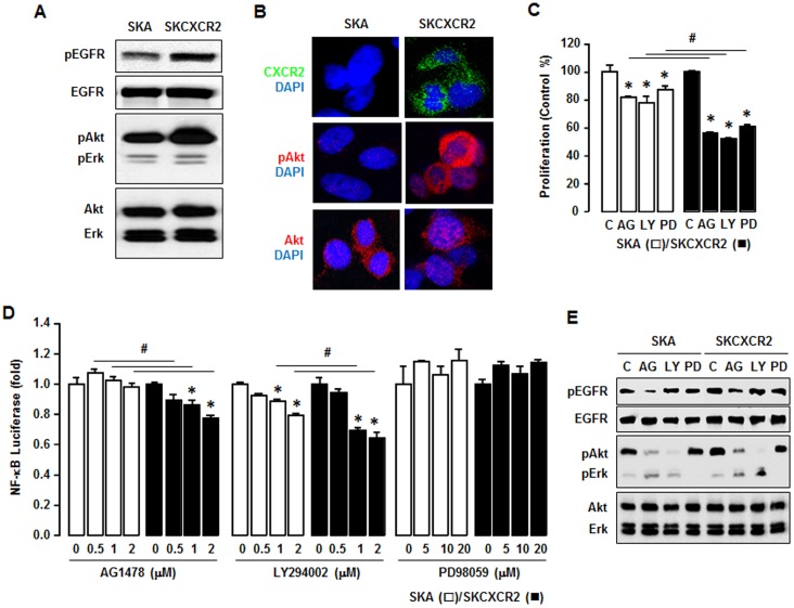
Figure 4. CXCR2 transactivates EGFR which contributes to NF-κB signaling via Akt activation.
(A) Comparison of EGFR activation in SKA and SKCXCR2 cells. Whole cell lysates were prepared and Western blots carried out using antibodies specific to EGFR, Akt, Erk and the phosphorylated forms (pEGFR, pAkt and pErk). The non-phosphorylated forms were used as loading controls. (B) Representative immunofluorescent staining patterns indicating Akt activation and CXCR2 protein expression levels in SKA and SKCXCR2 cells. (C) Comparative effects of AG-1478, LY294002 and PD98059 on cell proliferation in SKA and SKCXCR2 cells. Cells were incubated with vehicle (Control), AG-1478 (AG, 2 µM), LY294002 (LY, 2 µM) or PD98059 (PD, 20 µM) for 48 h. The cell proliferation assay was performed using MTT and values were normalized to untreated controls. * and # (p≤0.05) when compared to Controls (C) and SKA cells, respectively, by Student’s t-test. (D) Dose-dependent effects of EGFR downstream inhibitors on NF-κB luciferase activities in SKA and SKCXCR2 cells. After transfection with NF-κB luciferase vector overnight, cells were treated with AG-1478 (EGFR inhibitor, 0, 0.5, 1 and 2 µM), LY294002 (Akt inhibitor, 0, 0.5, 1 and 2 µM) or PD98059 (Erk inhibitor, 0, 5, 10 and 20 µM) for 4 h. * and # (p≤0.05) when compared to Controls (0 h) and SKA cells, respectively, by Student’s t-test. All experiments were performed at least in triplicate and data are shown as mean ± S.E. (E) Confirmation of specific inhibitors on EGFR, Akt and Erk activation in SKA and SKCXCR2 cells. Cells were treated with AG-1478 (2 µM), LY294002 (2 µM) and PD98059 (20 µM) for 4 h. Whole cell lysates were prepared and a western blot was carried out using antibodies specific to EGFR, Akt, Erk and their phosphorylated forms (pEGFR, pAkt and pErk). Non-phosphorylated forms were used as loading controls.

