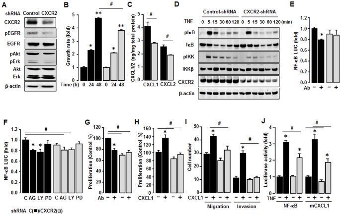
Figure 7. Inhibitory effect of CXCR2 shRNA on CXCR2-deriven cancer progression in SKCXCR2 cells.
(A) Knockdown of CXCR2 protein expression and comparison of EGFR activation after transfection of Control and CXCR2 shRNA in SKCXCR2 cells. After transfection with shRNAs for control and CXCR2 for 72 h, a Western blot was carried out using antibodies specific to CXCR2, EGFR, Akt, Erk and the phosphorylated forms (pEGFR, pAkt and pErk). The non-phosphorylated forms and β-actin were used as loading controls. (B) Comparison of growth rates in Control (black bars) and CXCR2 shRNA (gray bars) transfected SKCXCR2 cells. Cells were incubated for 0, 24 and 48 h and growth rates normalized to 0 h densities in each cell line. Experiments were performed in triplicate and all data are shown as mean ± S.E. * and ** (p≤0.05) in each group by ANOVA and Tukey’s pairwise comparisons. # (p≤0.05) between Control and CXCR2 shRNA transfected cells by Student’s t-test. (C) Cellular CXCL1 and CXCL2 concentrations in Control and CXCR2 shRNA transfected cells. Whole cell lysates were prepared, an ELISA carried out using antibodies specific to CXCL1 and CXCL2 and values normalized to total protein. (D) Effect of TNF (10 ng/ml) over time (0–120 min) on NF-κB activation in Control and CXCR2 shRNA transfected cells. Whole cell lysates were prepared and Western blots carried out using antibodies specific to IκB, IKK and their phosphorylated forms (pIκB and pIKK). β-actin was used as a loading control. (E) Effect of CXCL1/2/3 antibody on NF-κB luciferase activities in Control and CXCR2 shRNA transfected cells. After transfection of shRNA for 48 h followed by transfection of NF-κB luciferase vector overnight, cells were incubated with normal IgG and the CXCL1/2/3 antibody (Ab, 1∶100 dilution) for 4 h. (F) Effects of EGRF downstream inhibitors on NF-κB luciferase activity in Control and CXCR2 shRNA transfected cells. After transfection of shRNA for 48 h followed by transfection of NF-κB luciferase vector overnight, cells were treated with vehicle (C), AG-1478 (AG, 2 µM), LY294002 (LY, 2 µM) or PD98059 (PD, 20 µM) for 4 h. (G) Effect of CXCL1/2/3 antibody on cell proliferation in Control and CXCR2 shRNA transfected cells. Cells were incubated with normal IgG and CXCL1/2/3 antibody (Ab, 1∶100 dilution) for 48 h. The cell proliferation assay was performed using MTT and values were normalized to untreated controls. (H) Effect of CXCL1 on cell proliferation in Control and CXCR2 shRNA transfected cells for 48 h incubation. (I) Comparison of CXCL1-induced cell migration and invasion in Control and CXCR2 shRNA transfected cells. (J) Effect of TNF on NF-κB and mCXCL1 promoter luciferase activities in Control and CXCR2 shRNA transfected cells. After transfection of shRNA for 48 h followed by transfection of NF-κB or CXCL1 promoter luciferase vector overnight, cells were treated with TNF (10 ng/ml) for 4 h. All experiments were performed at least in triplicate and data are shown as mean ± S.E. * and # (p≤0.05) as calculated by Student’s t-test.

