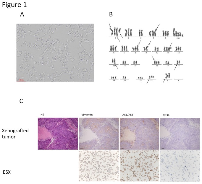
Figure 1. Establishment of the new epithelioid sarcoma cell line ESX.
A. Phase-contact microscopy findings for ESX.
B. Representative G-band karyotyping of ESX. The karyotype revealed 65~68, X, -X or –Y, add (X)(q22), +1, add(1) (p32), add(1)(q21), add(1)(q42), add(1)(q42), der(4;10)(q10;q10), add(8)(p11.2), -9, add(9)(p22), der(11)t(11;14)(p13;q13),-13, add(13)(q22), -14,-15,add(16)(p13.1), -17,-18, add(18)(q21),+21,add(22)(q13), +4~6mar[cp9]. Arrows indicate deletions and derivative chromosomes.
C. Immunostaining analysis of the xenografted tumors (scale bar, 100μm) and ESX cell line for vimentin and AE1/AE3, and CD34 (original magnification ×100). Subcutaneous inoculations of ESX cells into NOD/SCID mice produced growing tumors. Histologically, the xenografted tumors consisted of a distinct nodular arrangement of the tumor cells, a tendency to undergo central degeneration and necrosis, and an epithelioid appearance with cytoplasmic eosinophilia. The atypical cells were positive for vimentin and AE1/AE3, but negative for CD34.

