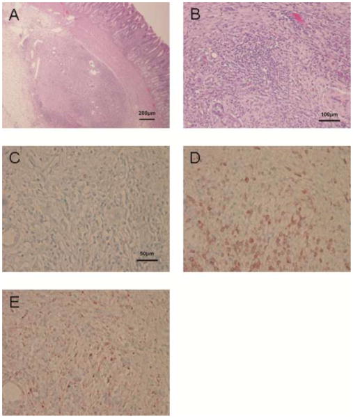
Figure 5.
Histopathological appearance of an area of severe cellular infiltration of remaining non-biopsied islets (at necropsy, in the same recipient pig as in Figure 4) 5 days after islet Tx into the GSMS. (A) Islet mass in the GSMS (H&E, x40). (B) Polymorphonuclear and mononuclear cell infiltrate in islet mass (H&E, x100). (C) Few insulin-positive beta cells were identified whenever severe cell infiltration of the islet mass was present. Islets were infiltrated particularly by (D) CD3+ and (E) CD68+ cells (x200). Similar histopathological findings were detected between biopsied islets (Figure 4) and remaining non-biopsied islets.

