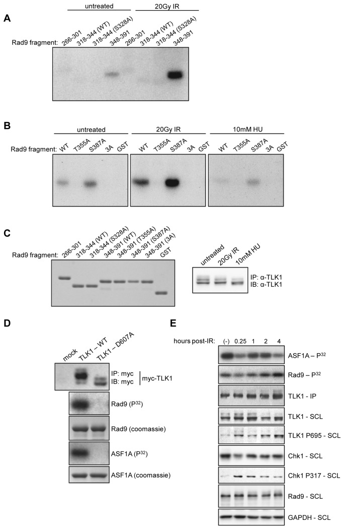
Figure 1. In vitro phosphorylation of Rad9 C-terminal fragments by TLK1.
A & B. Recombinant GST-fusion peptides corresponding to different regions of Rad9’s C-terminal tail were employed as substrates for in vitro kinase assays using TLK1 immunoprecipitated from HeLa cells exposed to the indicated conditions. 3A refers to a T355A/S363A/S387A triple mutant. C. Substrate input (Rad9, left panel) and kinase input (TLK1, right panel) for each kinase reaction. IP refers to immunoprecipitate, IB refers to immunoblot. D. Overexpressed, recombinant WT and kinase-dead (D607A) myc-TLK1 constructs were immunoprecipitated from HeLa cells and employed in in vitro kinase assays using full-length GST-Rad9 and GST-ASF1a as substrates. E. A time course in vitro kinase assay. TLK1 was immunoprecipitated from HeLa cells exposed to 20Gy IR and harvested at the indicated time-points. Immune complexes were incubated with either recombinant GST-Rad9 (amino acids 348-391) or GST- ASF1A. SCL refers to soluble cell lysates. Images shown are representative of two independent experiments.

