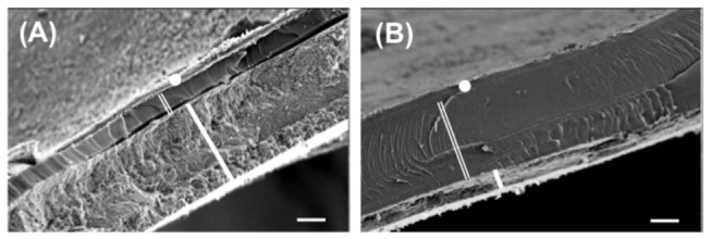
Figure 2. Scanning Electron Microscopy of the shell micro-scale structure of Notodiscus hookeri.

The cross sections show a layered architecture of the shell and two contrasted phenotypes. The outer periostracum (full white circle), the innermost mineralised layer (ML, thick white line) and, in between, an organic layer (OL, double white line) (A, B), the OL/ML ratio may be reversed according to snail population, BUS (A) or BRA (B). Scale bars, 10 µm.
