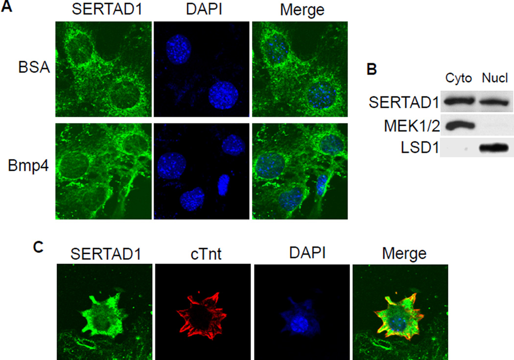
Figure 3. SERTAD1 is localized in both the cytoplasm and nucleus of cardiomyocytes.
(A) NkL-TAg cells were treated with BSA (negative control) or 100ng/ml BMP4 for 2 hours, followed by immunostaining using an anti-SERTAD1 antibody (green). The nuclei were visualized with DAPI staining (blue). The samples were examined under a Zeiss confocal microscope. Green signals were detected in both the cytoplasm and nucleus. No difference was observed between the BSA- and BMP4-treated samples. (B) The cytoplasmic and nuclear fractions of NkL-TAg cells were subjected to western analysis using a SERTAD1 antibody. MEK1/2 and LSD1 were used as cytoplasmic and nuclear controls, respectively. (C) Embryonic cardiomyocytes (E13.5) were isolated and cultured for 24 hours. Immunostaining was performed using antibodies against SERTAD1 (green) and cTnt (red). Total nuclei were visualized with DAPI staining (blue). Green signals were detected in both the cytoplasm and nucleus.

