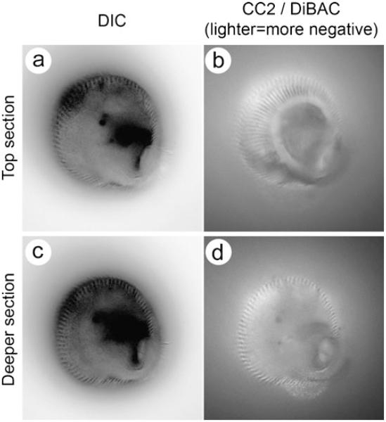
Fig. 10.
Multiple domains of membrane voltage within the single-celled ciliated protist Stentor coeruleus. An actively feeding Stentor was photographed at two different focal planes (a, b and c, d). In the differential interference contrast images left, the dark lines visible at the edges correspond to stripes of cilia that run along the cell from the oral to the aboral end. Right Images of the intricate and stable stripes of relatively hyperpolarized (lighter) and depolarized (darker) regions of the plasma membrane. The hyperpolarized stripes co-localize with the stripes of cilia. In d, the interior of the spiral feeding apparatus is visible as a spot of light surrounded by dark membrane. Despite the active phagocytosis of particles at this position on the membrane, this pattern is maintained. These fine scale differences in voltage are highly stable

