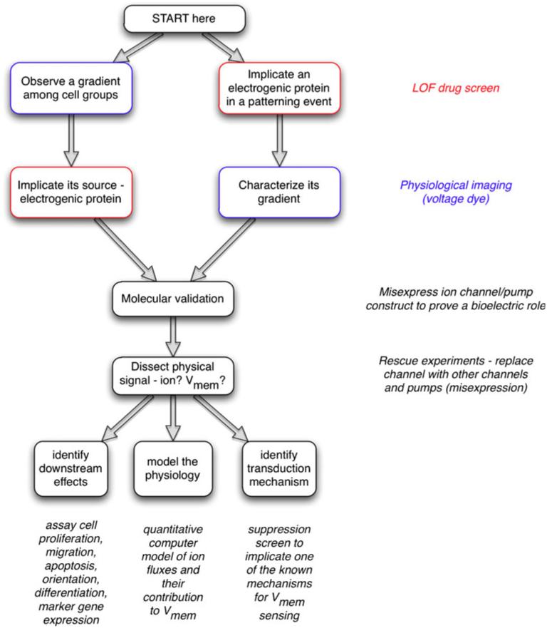
Fig. 2.
Strategy for investigating bioelectric signaling. One often begins by detecting an interesting bioelectrical gradient during some process and hypothesizing that this voltage or ion flow pattern is functionally important. In this case, the next step is to develop a robust assay recapitulating this process and to conduct a loss-of-function hierarchical drug screen to narrow down a list of candidate transporters that might underlie the observed gradient. In contrast, a specific transporter candidate might be suggested by the results of a genetic screen, network analysis, or microarray comparison. Because Vmem is determined by ion flux, part of characterizing bioelectric signals is determining the endogenous translocators involved. Thus, the next step would involve the use of fluorescent voltage- or ion-reporting dyes (with and without a blocker of the channel/pump) to characterize the spatiotemporal properties of whatever gradient pattern might be produced by the transporter in question (Adams and Levin 2012a, 2012b; Oviedo et al. 2008). Molecular-genetic validation of the pharmacological data follows; a loss-of-function experiment with the knockout or misexpression of a dominant negative mutant should be used to establish that this transporter is indeed important for the phenotype under study. Next, to determine the physical process carrying instructive information, the transporter is blocked and a rescue of the phenotype is attempted with other transporters that distinguish among non-ionic, ion-specific and voltage-specific functions. Subsequently, this biophysical signal has to be connected to canonical pathways by (1) identifying the second-messenger mechanism(s) by which a biophysical signal is transduced into changes in cell behaviors, (2) identifying its downstream effects (which genes does it turn on/off, how does it affect cell behavior) and (3) modeling the physiology (quantitative spatial simulation of the ion flows that result in the relevant voltage gradient)

