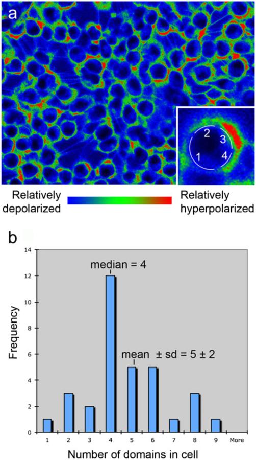
Fig. 8.
Multiple domains of membrane voltage within single cells in culture. a Monolayer of cultured COS M6 cells were imaged by using the membrane potential reporters DiBAC4(3) and CC2-DMPE (red the most negatively charged membrane, blue the least negatively charged membrane). A domain was defined by coloring the original black and white image by using a red/green/blue lookup table in IPLabs, the software used to collect the images. The image was then opened in Photoshop and any stretch of color in which the pixels were pure red, pure green, or pure blue were counted as a region. Thus, this count reveals three possible values of membrane voltage. Inset Higher magnification of a single cell showing that this cell was determined to have four domains. b Domains were counted on 33 randomly chosen cells. These cells had 5±2 (mean±SD) domains per circumference; the median number of domains was four. Thus, even on using a conservative counting protocol, these cells were found to maintain four to five voltage domains around the circumference of the cell within the plane of focus

