
Figure 1.
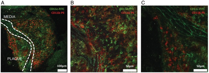
Localization and characterization of CD11c+ cells in atherosclerotic plaques of the aortic root of Apoe−/−CD11cDNR mice fed a normal chow diet for 20 weeks. (A–C) Multiphoton microscopy. CD11c+ (FITC, green) and CD11b+ (PE, red) cells are present in the plaque and lymphoid aggregates in the adventitia. White lines indicate the media. (B) Detail of the adventitia demonstrating infiltration of CD11c+ cells (FITC). (C) Detail of the atherosclerotic plaque. CD11c+ cells (FITC) and CD11b+ cells (PE) are present in the shoulder region of the plaque.