
Fig. 3.
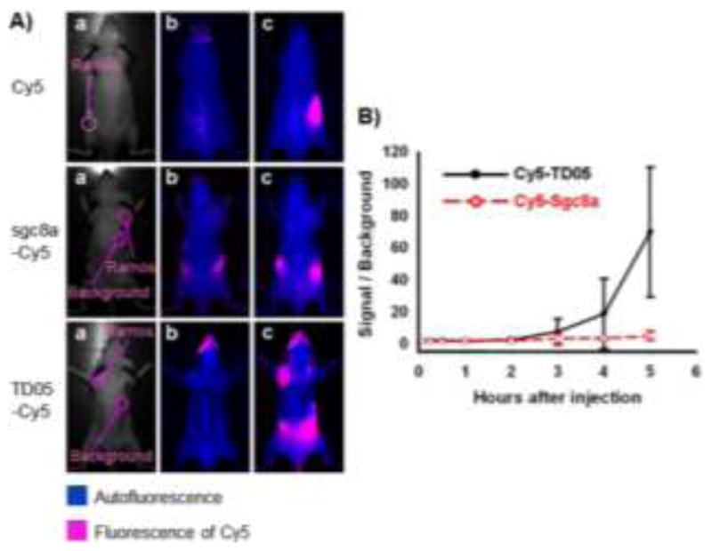
An aptamer identified through cell-SELEX for in vivo imaging of Ramos subcutaneous xenograft. (A) Optical and fluorescent imaging of tumors in mice using Cy5 dye and Cy5-labeled aptamers. Target Ramos cells were injected subcutaneously on the back of BALB/c nude mice. Cy5 dye, Cy5-labeled sgc8a (control), or Cy5-labeled TD05 was subsequently injected intravenously, and images of the dorsal side of live mice were taken at various time points after injection. Images a, b and c represent the optical photographs, the fluorescent images before injection, and 3.5 h after injection, respectively. (B) Quantification of the signal-to-background ratio for Cy5-labeled TD05 and Cy5-labeled sgc8a. The number of mice used to derive statistical information for each probe is 5. Data represent mean ± standard error. Adapted from ref.54