Figure 1.
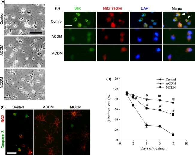
ACDM and MCDM protect OPCs against growth factor withdrawal-induced apoptosis, as well as support long-term OL survival. (A) Representative phase contrast micrographs show that OPCs maintained in the control medium (without growth factors) started to degenerate at 48 h following growth factor withdrawal, while OPCs maintained in ACDM or MCDM remained intact and viable. (B) Triple-labeling of OPCs with Bax, MitoTracker, and DAPI revealed that at 24 h, many cells in the control showed typical Bax translocation from cytosol to mitochondrial membrane (arrowheads), which were not observed in cells cultured in ACDM or MCDM. (C) Numerous OPCs in the control medium showed positive immunostaining of activated caspase-3, while very few, if any, were found in ACDM or MCDM-treated cultures, 24 h following the exposure. (D) Although both ACDM and MCDM could support long-term OL survival, ACDM was significantly more effective than MCDM after 48 h. #P < 0.01 versus control; *P < 0.01 versus MCDM. Data are mean ± SEM from three independent experiments. Scale bars: 50 μm.

