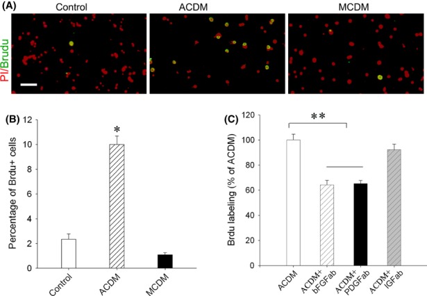
Figure 2.

ACDM, but not MCDM, promotes OPC proliferation. After being exposed to the control or the conditioned medium for 48 h, OPC proliferation was assessed by BrdU labeling. (A) Representative photographs show that the number of BrdU+ cells, which was minimal in the control and MCDM group, was significantly higher in ACDM-treated cultures. (B) Quantification of cell proliferation by calculating the percentage of BrdU+ to total cells (PI counterstained nuclei). (C) Blocking PDGFaa and bFGF, but not IGF-1, by their specific neutralizing antibodies, significantly reduced ACDM-enhanced OPC proliferation. *P < 0.01 versus MCDM and the control, and **P < 0.01 versus ACDM. Data are mean ± SEM from three independent tests. Scale bar: 100 μm.
