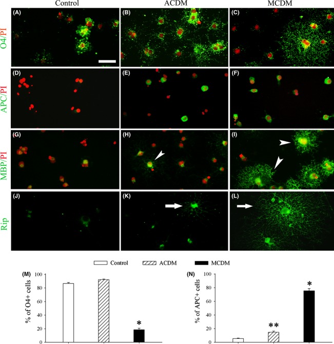
Figure 3.
MCDM, but not ACDM, strongly enhances OL differentiation. (A–L) Representative photographs show the effects of ACDM and MCDM on OL differentiation, as assessed using a panel of antibodies against developmental stage-specific OL markers (see text for details), 8 days after the exposure. Most cells in the control medium degenerated as evidenced by pyknotic nuclei (red, PI labeled), while few survival cells remained undifferentiated as indicated by O4+ (A), APC- (D), and MBP- (G) phenotype. Although ACDM greatly enhanced OL survival, it had only weak effect of OL differentiation, indicated by high percentage of O4+ cells (B) and lower percentage of APC+ cells (E), as well as weakly immunostained MBP that was mostly detected in cell somata (an arrowhead in H). In contrast, MCDM-exposed cultures had fewer O4+ (C) but more APC+ cells (F), and the differentiated cells were intensively labeled with MBP in both somata and processes (arrowheads in I). Rip immunostaining revealed fine processes in MCDM-exposed (arrow in L) versus ACDM-exposed (arrow in K) or the control (J) cultures. Scale bar: 50 μm. Quantification of O4+ and APC+ cell number are shown in (M) and (N), respectively. *P < 0.01 versus control or ACDM; **P < 0.05 versus control.

