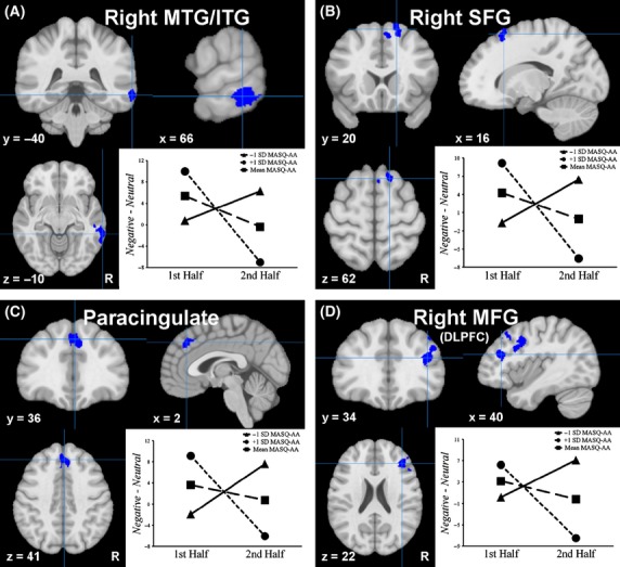
Figure 2.

Moderation of habituation to negative stimuli by anxious arousal. SFG, superior frontal gyrus; MFG, middle frontal gyrus; DLPFC, dorsolateral prefrontal cortex; MTG, middle temporal gyrus; ITG, inferior temporal gyrus; Blue, high MASQ-AA associated with habituation. The graphs depict the change in neural response to negative words over time at +1 (circle endpoints) and −1 (triangle endpoints) standard deviations (SD) and the mean (square endpoints) of the Anxious Arousal subscale of the Mood and Anxiety Symptom Questionnaire (MASQ-AA). Time (1st task half and 2nd task half) is plotted on the x-axis against brain activation related to negative stimuli (negative minus neutral) on the y-axis. Graphs reflect values with normalized PSWQ and MASQ-AD-LI partialled out.
