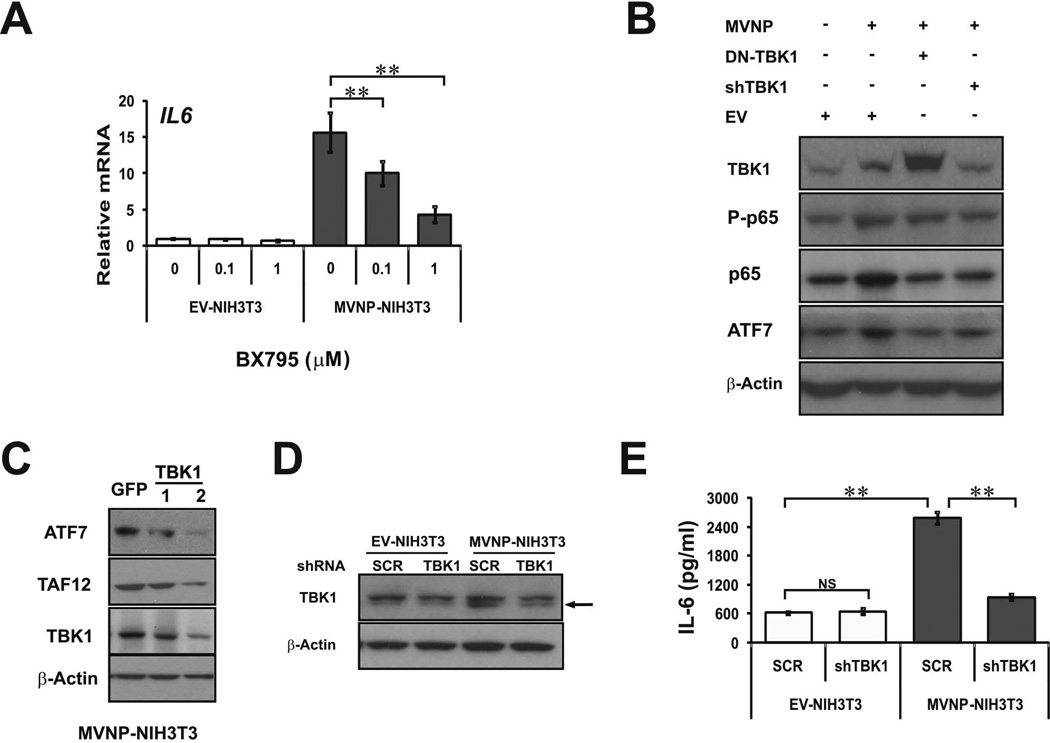
Figure 4. Inhibition or knockdown of TBK1 in NIH3T3 and HEK293 cells impaired MVNP effects.
(A) EV-NIH3T3 and MVNP-NIH3T3 cells were treated with TBK1 inhibitor BX795 (0, 0.1, 1 µM) for 24 hrs and endogenous IL6 RNA expression was analyzed by qPCR. (B) Lysates from HEK293 cells transfected with the following vectors for 36 h: EV, MVNP alone, 1:1 MVNP:TBK1(K38A) (DN-TBK1), or MVNP plus shTBK1 plasmid were immunoblotted for TBK1, total and phosphorylated-p65 NF-κB (P-p65), ATF7, and β-actin. (C) Lysates from MVNP-NIH3T3 cells infected with 2 different shTBK1 lentiviruses were immunoblotted for ATF7, TAF12, TBK1 and β-actin. (D, E) EV- and MVNP-NIH3T3 cells infected with scrambled (SCR) control and shTBK1 lentiviruses were treated with puromycin (2 µg/mL) at 6 d post-infection and thereafter. (D) At 6 d post-infection, cell lysates were immunoblotted for TBK1 protein (lower band is TBK1; upper band is non-specific) and β-actin, and (E) secreted IL-6 levels in cell culture supernatants were detected by ELISA assay at 38 d post-infection. NS, not significant; ** (ρ<0.01), significantly different between samples as indicated.

