Figure 4.
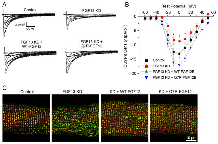
Both WT and Q7R-FGF12 rescue Ca2+ current density and localization from FGF13 KD. A: Representative Ca2+ current traces from the 4 groups. B: I–V curve depicting the rescue of CaV1.2 current density with WT and Q7R-FGF12. Peak current densities at 0 mV are −13.01 ± 0.88 pA/pF (n = 9) for control, −8.09 ± 1.30 pA/pF (n = 13) for FGF13 KD, −12.86 ± 1.50 pA/pF (n = 13) for FGF13 KD + WT-FGF12, and −17.05 ± 1.46 (n = 13) for FGF13 KD + Q7R-FGF12. C: Immunostaining for CaV1.2 (green) and ryanodine receptor (red) showing that CaV1.2 is mislocalized with FGF13 KD and the localization is rescued with WT and Q7R-FGF12. *P < .05 versus control. FGF = fibroblast growth factor; KD = knocked down; WT = wild type.

