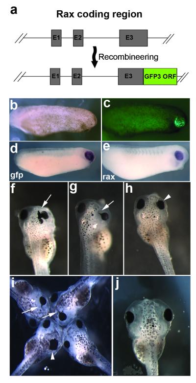
FIG. 2.
Expression of the rax-gfp3 fusion BAC. (a) Detail of rax coding region contained in BAC 349A23, showing construction of the rax GFP3 fusion construct. (b) Bright field view of stage 24 embryo injected with rax-gfp fusion BAC. (c) GFP fluorescent view of same embryo, showing expression of transgene in developing retina. (d) In situ hybridization against gfp3 mRNA in BAC-injected, stage 24 embryo. (e) In situ hybridization of WT sibling embryo showing endogenous rax mRNA expression. (f-h) rax overexpression phenotypes seen in stage 42-43 tadpoles injected with rax-gfp fusion BAC. Arrows indicate formation of ectopic retinal pigmented epithelium. Arrowheads indicate eyes shifted medially. (i) rax overexpression phenotypes observed in rax mRNA injected embryos. Arrows and arrowheads indicate the same structures as in f-h. (j) Normal, wildtype stage 42 embryo for comparison.

