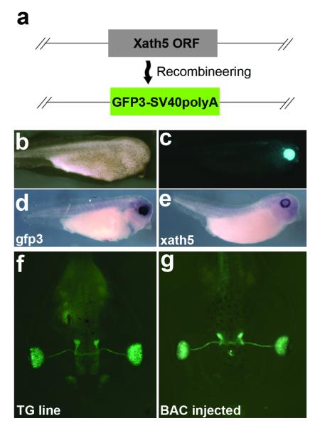
FIG. 3.
Expression of the xath5 GFP reporter BAC. (a) Detail of xath5 coding region contained in BAC 38N10, showing replacement of coding region with gfp3 open reading frame (ORF). (b) Bright field image of stage 35 embryo injected with xath5 reporter BAC. (c) gfp fluorescence image of same embryo, showing gfp3 expression in developing retina. (d) In situ hybridization against gfp3 mRNA in BAC-injected, stage 35 embryo. (e) In situ hybridization of WT sibling embryo showing endogenous xath5 mRNA expression. (f) Stage 46 F2 tadpole from integrated line, using xath5 promoter to drive GFP expression. (g) Stage 46 xath5 BAC-injected tadpole GFP expression is comparable to integrated line expression. Embryos were PTU treated to visualize retinal tissue (b-g).

