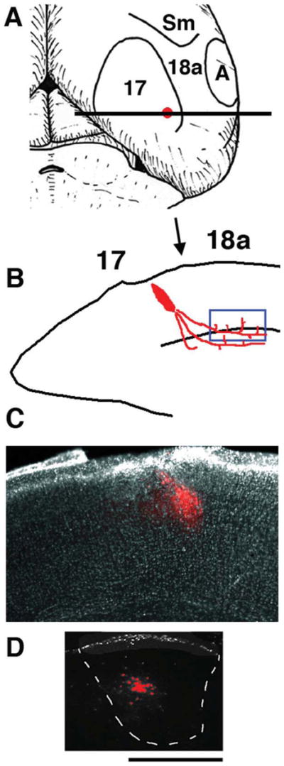
Figure 1.

Tracer injections into area 17 to reveal anterogradely labeled axons projecting from area 17 to ipsilateral area 18a. Medial is to the left. A: Diagram of top view of right hemisphere showing areas 17 and 18a and approximate location of the intra-cortical injection of anterogradely transported fluorescent tracers dextran Alexa into area 17. B: Diagram of coronal tissue section taken at the level of the horizontal bar in A illustrating the tracer injection in area 17 and the location of the labeled fibers in area 18a analyzed with time-lapse methods (blue box near white matter). Arrow indicates presumptive 17/18a border. C: Low-power image of coronal tissue section showing an injection of Alexa 594 into area 17. D: Image from a coronal section showing the anterogradely labeled field observed in the ipsilateral dLGN following the Alexa 594 injection shown in C. Scale bar = 500 μm.
