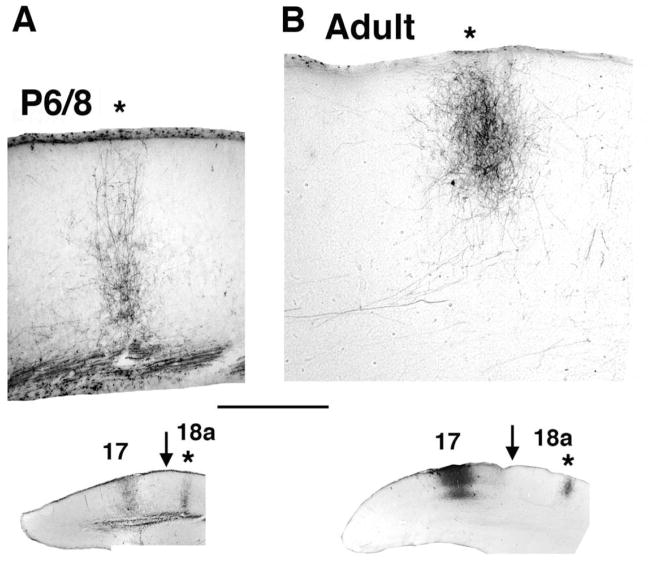
Figure 3.
Area 17–18a projections at P8 and adulthood. A, B: Images from coronal tissue sections illustrating the area 17–18a projections labeled following discrete intracortical injections of the anterogradely transported tracer BDA into area 17. Medial is to the left. A: Column-like field of BDA-labeled fibers in area 18a in an animal (case L24A) injected with BDA into area 17 at P6 and studied at P8. B: Column-like field of BDA-labeled fibers in area 18a in an adult animal (case SI4). Locations of the injection sites in area 17 and the labeled fields in area 18a (asterisks) are shown in the lower insets. Arrows in insets indicate approximate location of 17/18a border. Scale bar = 500 μm.

