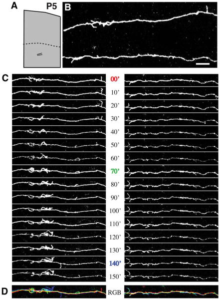
Figure 5.
Axons in the subcortical white matter extend numerous, dynamic interstital filopodia. A: At P5, the boundary between gray matter and white matter (dotted line) is roughly 700 μm below the pial surface. B: Segments from a pair of axons running roughly parallel to the pial surface beneath area 18a were imaged by two-photon time-lapse microscopy in a living cortical slice made from P5 rat brain. The location of the pair of imaged axons is schematized in the low-magnification reconstruction in A. C: Images collected at 10-minute intervals reveal highly motile interstitial filopodia. D: Temporal overlay of three time points from series in C. Images from each time point (0, 70, and 140 min) are different colors, and white represents stable regions of the axon. Scale bar = 10 μm.

