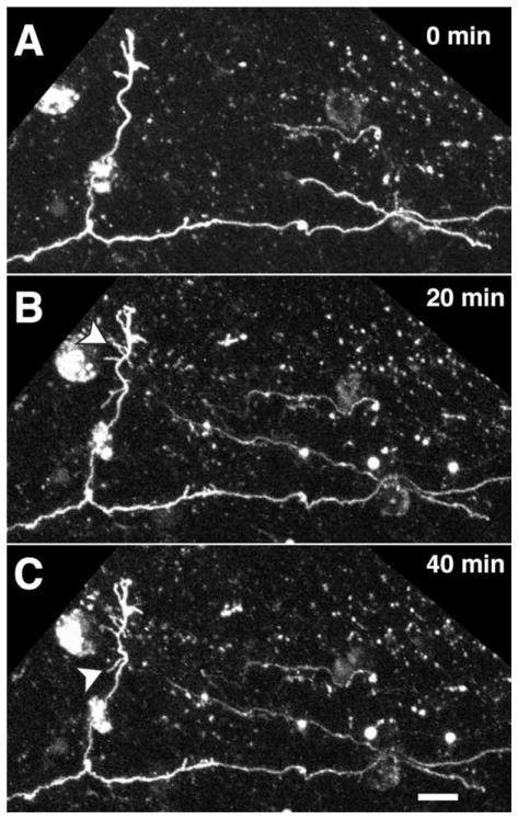
Figure 7.
Pial-directed interstitial branch formed in the white matter along a parental axon from area 17 in a P6 rat pup. A–C: Series of images showing morphological rearrangements (arrowheads) at 20-minute intervals reveals a high degree of filopodial exploration near the tip of the single large interstitial branch that has invaded cortical gray matter. Relatively little filopodial activity is observed along the parental axon at the same times. Scale bar = 10 μm.

