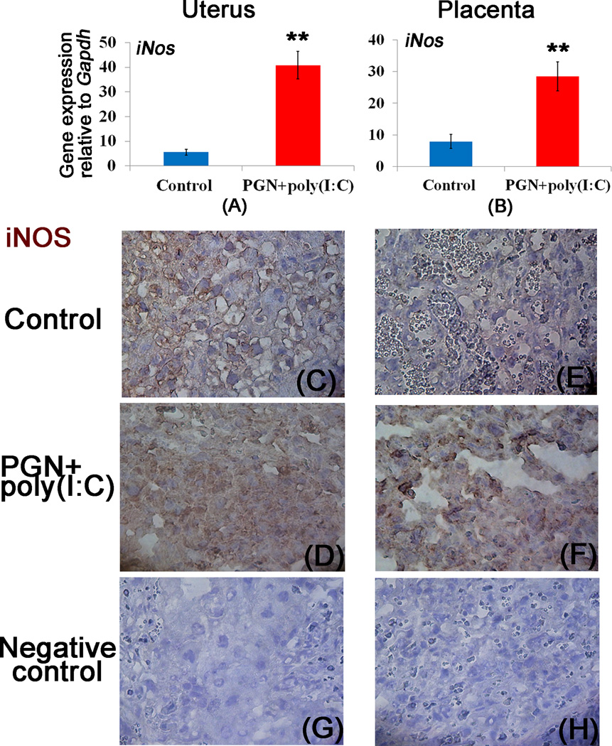
Figure 2. Decrease of a2V is associated with induction of iNOS.
Panels A and B show mRNA expression of iNOS; panels C–D and E–F show immunolocalization of iNOS in the uterus and placenta recovered from the control and PGN+poly(I:C) treated groups, respectively. Panels G and H show the staining with isotype control antibodies for mouse IgG and rabbit IgG respectively. N=6–11 each group. Error bars= ±SEM. **P≤0.01 Significant difference vs respective control. Six sections per animal were analyzed. Original magnification: 400X.

