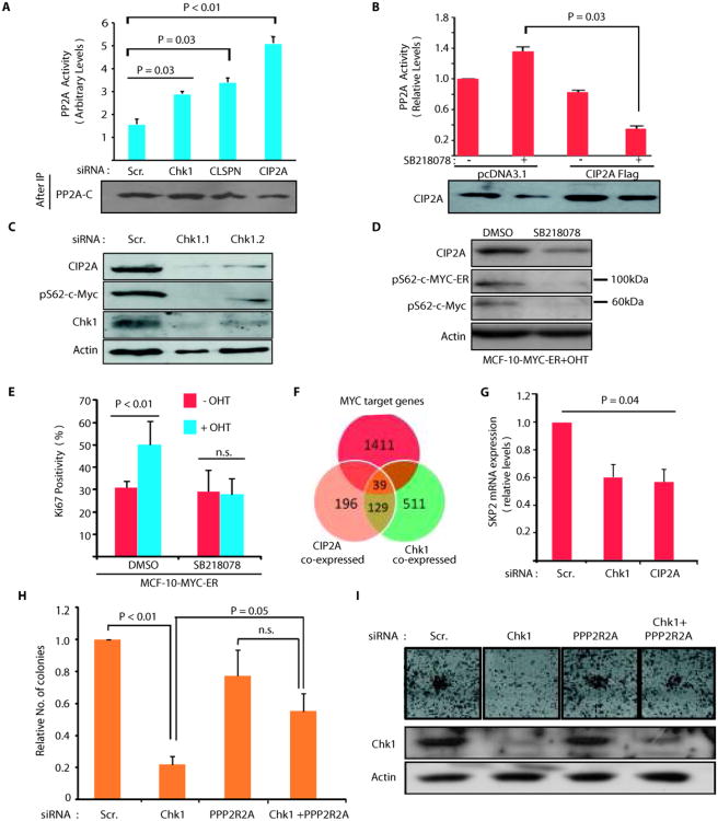
Figure 5.
Chk1 inhibition re-activates PP2A tumor suppressor activity. A, Depletion of Chk1, Claspin or CIP2A induces PP2A activity in AGS cells. B, PP2A activity in pcDNA3.1 or CIP2A Flag transfected AGS cells treated with SB218078 (1uM; 48h) as indicated. C, Chk1 depletion inhibits CIP2A protein expression and of serine62 phosphorylated MYC in AGS cells 72h post transfection. D, Western blot analysis of SB218078 effects(1 uM, 24h) on serine62 phosphorylation of MYC-ER fusion protein (appr. 100 kDa) or endogenous MYC protein (appr. 65 kDa) in tamoxifen-treated MCF-10 cells stably transfected with MYC-ER. E, Serum starved MYC-ER expressing MCF-10A cells, pretreated for 24h with either DMSO or SB218078(1 uM), were induced to MYC-mediated proliferation by tamoxifen treatment. Quantitation of Ki-67 positive cells demonstrates requirement of Chk1 activity for MYC-induced proliferation. F, Venn diagram displaying overlap of genes that significantly associate with either CIP2A (orange) or Chk1 (green) or MYC (red) expression in human neuroblastomas (n= 168), p < 10−10, hypergeometric distribution. G, Real-time PCR analysis of SKP2 mRNA expression from AGS cells transfected with CIP2A and CHk1 siRNA for 72 hours. H-I, Inhibition of CIP2A target PP2A B-subunit PPP2R2A expression by siRNA rescues Chk1 siRNA effects on the AGS cell colony growth. Shown are mean values + S.D., of representative results from three independent experiments (student's t-test).

