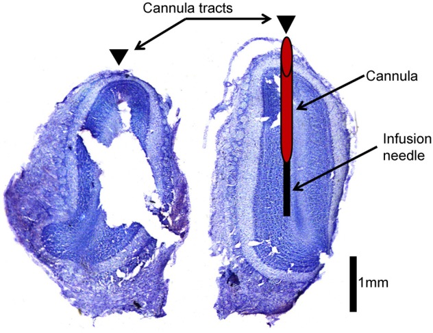
Figure 2.

Histological verification of cannula placement. This is an example of an olfactory bulb slice verifying cannula placement. Arrowheads indicate location of cannula tracts. The left hemisphere indicates where cannulae were implanted (red cylinder) and how far the infusion needle extends beyond the cannula (black rectangle) during infusion of drug into the olfactory bulb.
