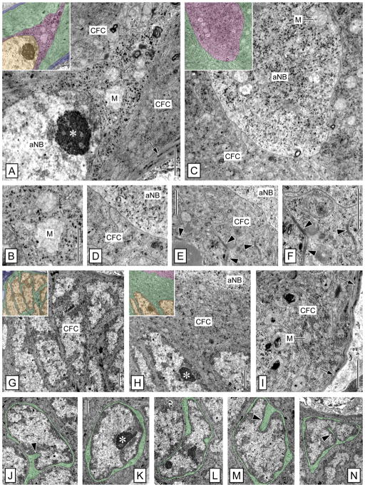
Figure 7.
Ultrastructure of the clump of cells: Clump-forming cells and adult neuroblast. TEM micrographs of cross-sections through the clump of cells in the lateral soma cluster. Colorized insets highlight different cell types: adult neuroblast (magenta), clump-forming cells (green), processes of cell body glia (blue), nuclei of all cell types (yellow). A,B: Peripheral domain of the adult neuroblast (aNB) at the apex of the clump of cells. The electron-lucent cytoplasm of the peripheral domain of the aNB contains large electron-lucent mitochondria (M) and numerous electron-dense granules. The peripheral domain of the aNB is completely covered by outer processes of clump-forming cells (CFC) whose cytoplasm differs distinctly from that of the aNB in electron density and organelle composition (arrow, processes of cell body glia; asterisk, nucleolus). C–F: Bulbous foot of the adult neuroblast (aNB) in the nucleus-free center of the clump of cells. The bulbous foot of the aNB is identical to the peripheral domain in having electron-lucent cytoplasm containing large electron-lucent mitochondria (M) and numerous electron-dense granules. The bulbous foot of the aNB is completely covered by the inner processes of clump-forming cells (CFC). No membrane specializations are apparent at the interface between aNB and CFCs. Occasional desmosome-like membrane specializations are present between adjacent processes of different CFCs (arrowheads). G–N: Ultrastructure of clump-forming cells (CFC). G: Cortex of the clump of cells. The cortex of the clump of cells is formed by a continuous layer of CFC somata characterized by a relatively large and irregularly shaped nucleus and only a thin rim of cytoplasm. H,I: Nucleus-free center of the clump of cells. The nucleus-free center of the clump of cells is filled by the bulbous foot of the aNB and a thick layer of inner processes of clump-forming cells (CFC) surrounding it. The cytoplasm of the CFC processes is of medium electron density and contains small mitochondria (M) and delicate cisternae of rough ER (arrows, processes of cell body glia; asterisk, nucleolus). J–N: Somata of clump-forming cells with cytoplasm colorized green to highlight the irregular and diverse shape of somata and nuclei as well as the high nuclear:cytoplasmic ratio of CFCs. Note deep invaginations present in some CFC nuclei (arrowheads). Asterisk, nucleolus. Scale bars = 1 μm A (applies to A,B); 1 μm in B (applies to B,D); 1 μm in E–I; 1 μm in K (applies to J–N).

