
FIGURE 2. Doxorubicin induces caspase-1 independent processing and release of IL-1β in LPS-primed BMDC.
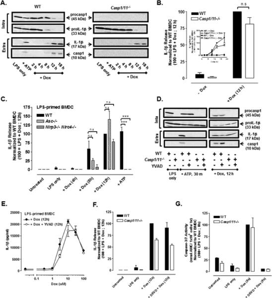
(A) LPS-primed (1 μg/ml) WT or Casp1/11−/− BMDC were stimulated with Dox (10 μM) for 2-18 h, and western blot analysis of IL-1β and caspase-1 from cell lysates and extracellular supernatants was performed. BMDC were LPS-primed for 5.5 h followed by 30 min of ATP (5mM) stimulation. Results are representative of 3 identical experiments. (B) The release of IL-1β from LPS-primed (1ug/ml) WT or Casp1/11−/− BMDC stimulated or not with 10 μM Dox for 12 h was assayed by ELISA. IL-1β release was normalized to WT BMDC treated with LPS + Dox for 12 h and expressed as the mean ± SE of 5 experiments. The differences between WT and Casp1/11−/− BMDC were not significant (n.s, P > .05) by Student's t-test. Inset: Kinetics of Dox-induced IL-1β release (normalized to WT BMDC treated with LPS + Dox for 12 h) in LPS-primed WT versus Casp1/11−/− BMDC as described in part (A). Results are the mean ± SE of 3 experiments. (C) WT, Asc−/−, and Nlrp3−/− Nlrc4−/− BMDC were primed with LPS for 4 h before stimulation with Dox for 4, 8, or 12 h and assayed for release IL-1β release by ELISA. Parallel samples were primed with LPS for 15.5 h prior to ATP (5mM) stimulation for 30 min. IL-1β release was normalized to WT BMDC treated with LPS + Dox for 12 h and expressed as the mean ± SE of 3-5 experiments. ***P < .001 or n.s by ANOVA. (D) Western blot analysis of LPS-primed (1μg/ml) WT or Casp1/11−/− BMDC stimulated with 10 μM Dox for 12 h or 5mM ATP for 30 min in the presence or absence of the caspase-1 inhibitor, YVAD (50 μM). BMDC were LPS-primed for 7.5 h followed by ATP (5mM) stimulation for 30 min. The data are representative of results from 2 experiments. (E) Concentration-response relationship for Dox-stimulated IL-1β release in the presence or absence of YVAD in LPS-primed BMDC. The curve depicted without YVAD treatment is the same as shown in Figure 1D. Results are the mean ± SE of 3 experiments. (F) The release of IL-1β was measured in LPS-primed WT or Casp1/11−/− BMDC stimulated ± 10μM Dox for 12 h in the presence or absence of DEVD (50μM). IL-1β release was normalized to WT BMDC treated with LPS + Dox for 12 h. Results are the mean ± range of 2 experiments. (G) Caspase-3/7 activity was measured in LPS-primed WT and Casp1/11−/− BMDC ± 10 μM Dox in the presence or absence of DEVD. Casp3/7 activity was normalized to WT BMDC treated with LPS + Dox for 8 h. Results are the mean ± range of 2 experiments.