
FIGURE 3. TLR4 activation coupled with caspase-8 inhibition induces RIP1/RIP3-dependent necroptosis and release of unprocessed proIL-1β while TLR4 activation coupled with Dox treatment induces caspase-8-dependent apoptosis.
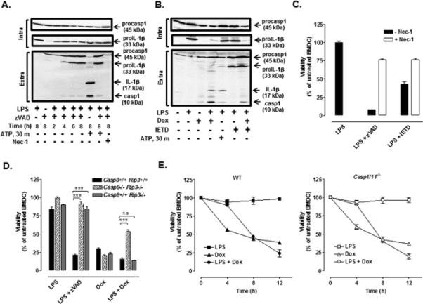
(A) Western blot analysis of IL-1β and caspase-1 in cell lysates and extracellular medium from WT BMDC treated with LPS (1μg/ml) and zVAD (50 μM) for 2-8 h with or without necrostatin-1 (Nec-1, 50 μM). (B) Western blot analysis of IL-1β and caspase-1 in cell lysates and extracellular medium from WT BMDC treated with LPS for 4 h prior to stimulation with Dox (10 μM) in the presence or absence of IETD (100uM) for 12 h. In panels A and B, BMDC were primed with LPS for 7.5 h prior to 30 min of ATP (5mM) stimulation. The data are representative of results from 2-3 experiments. (C) WT BMDC were co-treated with LPS + zVAD or LPS + IETD in the presence or absence of Nec-1, and cell viability was assessed by measuring intracellular ATP content. Results are from a single experiment with each condition performed in quadruplicate. (D) WT, Casp8−/− Rip3−/−, and Casp8+/+ Rip3−/− BMDC were treated with LPS (16 h), Dox (12h), or LPS for 4 h prior to Dox or zVAD for 12 h, and cell viability was assessed. Results are from a representative experiment (of 2 similar experiments) performed in quadruplicate. ***P < .001 by ANOVA. (E) WT and Casp1/11−/− BMDC were LPS-primed or not for 4 h prior to stimulation or not with Dox for 4, 8, or 12 h, and cell viability was assessed. Results are from a representative experiment of 2 similar experiments with each condition performed in triplicate.