
FIGURE 4. Doxorubicin induces caspase-8 dependent IL-1β processing and release in LPS-primed BMDC.
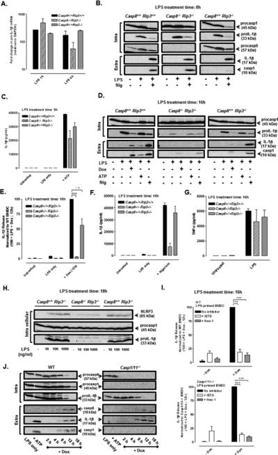
(A) ProIL-1β mRNA (normalized to GAPDH) was measured by qPCR in WT, Casp8−/− Rip3−/−, and Casp8+/+ Rip3−/− BMDC treated with LPS (1 μg/ml) for 1 h or 4 h. Results are from a single experiment. (B) Western blot analysis of proIL-1β, procaspase-1, and procaspase-8 in cell lysates (intra) and mature IL-1β and caspase-1 p10 subunit in extracellular (extra) supernatants of WT, Casp8−/− Rip3−/−, and Casp8+/+ Rip3−/− BMDC treated with LPS for 8 h and stimulated ± nigericin (10 μM) for the final 30 min of the LPS treatment period. Data are representative of results from 2 experiments. (C) BMDC of the indicated genotypes were treated with LPS (1 μg/ml) for 7.5 h prior to simulation ± ATP (5 mM) for an additional 30 min; IL-1β release was assayed by ELISA. Results are the mean ± range of 2 experiments. (D) Western blot analysis of pro-IL-1β and procaspase-1 in cell lysates (intra) and mature IL-1β and caspase-1 p10 subunit in extracellular (extra) supernatants of WT, Casp8−/− Rip3−/−, and Casp8+/+ Rip3−/− BMDC treated with LPS for a total of 16 h and stimulated with either Dox (10 μM) for the final12 h, or with ATP (5 mM) or nigericin (10 μM) for the final 30 min, of the LPS treatment period . Results are representative of 3 similar experiments. (E, F) WT, Casp8−/− Rip3−/−, and Casp8+/+ Rip3−/− BMDC were treated with LPS (1 μg/ml) for a total of 16 h and stimulated with Dox (10 μM) for the last 12 h (E) or nigericin (10 μM) for the last 30 min (F) of the LPS treatment period. IL-1β release was assayed by ELISA and normalized to WT stimulated with LPS + Dox (E) or WT stimulated with LPS + nigericin (F). Results are the mean ± SE of 3 experiments for (E) or the mean ± SE for 2 experiments (F) ***P < .001 or not significant (n.s) by ANOVA. (G) TNFα release (by ELISA) from WT, Casp8−/− Rip3−/−, and Casp8+/+ Rip3−/− BMDC treated ± LPS (1 μg/ml) for 16 h. Results are the mean ± range of 2 experiments. (H) Western blot analysis of proIL-1β, procaspase-1, and NLRP3 expression in WT, Casp8−/− Rip3−/−, and Casp8+/+ Rip3−/− BMDC treated for 18 h with 0, 10, 100, or 1000 ng/ml of LPS. Results are representative of 2 experiments. (I) WT and Casp1/11−/− BMDC were treated with LPS (1 μg/ ml) for a total of 16 h and stimulated ± Dox (10 μM), ± IETD (100 μM), ± Nec-1 (50 μM) for the final 12 h of the LPS treatment period. IL-1β release was assayed by ELISA and normalized to the samples stimulated with LPS + Dox. Results are the mean ± SE of 3 experiments. ***P < .001 by ANOVA. (J) Western blot analysis of procapase-8, procaspase-1, and proIL-1β in cell lysates (intra) and mature IL-1β, caspase-8 p18 subunit, and caspase-1 p10 subunit in the extracellular supernatants (extra) from WT or Casp1/11−/− BMDC treated with LPS (1 μg/ml) for 4 h prior to co-stimulation with Dox (10 μM) for another 2-18 h or with LPS for 5.5 h prior to ATP (5mM) stimulation for 30 min. The data are representative of results from 3 experiments.