
FIGURE 6. Staurosporine induces caspase-8-dependent IL-1β processing and release in LPS-primed BMDC.
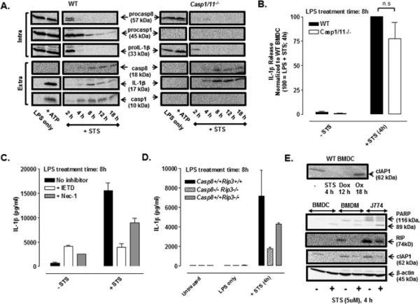
(A) Western blot analysis of procapase-8, procaspase-1, and proIL-1β in cell lysates (intra) and mature IL-1β, caspase-8 p18 subunit, and caspase-1 p10 subunit in the extracellular supernatants (extra) from WT or Casp1/11−/− BMDC were treated ± LPS (1 μg/ml) for 4 h and then co-stimulated with STS (5 μM) for an additional 2-18 h or with LPS for 5.5 h prior to ATP (5mM) stimulation for 30 min. The data are representative of results from 3 experiments. (B) LPS-primed (1 μg/ml, 4h) WT and Casp1/11−/− BMDC were stimulated ± STS (5 μM) for 4 h; IL-1β release was assayed by ELISA and normalized to WT BMDC treated with LPS + STS (4 h). Results are the mean ± SE of 4-5 experiments. The differences between WT and Casp1/11−/− BMDC were not significant (n.s, P > .05) by Student's t-test. (C) WT BMDC were primed with LPS (1 μg/ml, 4 h) prior to stimulation ± STS (5 μM, 4 h) in the presence or absence of IETD (100 μM) or Nec-1 (50 μM); IL-1β release was assayed by ELISA. Results are from a single representative experiment of 3 experiments. (D) WT, Casp8−/− Rip3−/−, and Casp8+/+ Rip3−/− BMDC were treated ± LPS (1 μg/ml) for 4 h prior to co-stimulation ± STS (5 μM) for an additional 4 h. Results are from a single experiment with each condition was performed in duplicate. (E) Upper panel: Western blot analysis of cIAP1 of lysates from untreated WT BMDC or WT BMDC treated with STS (5 μM, 4 h), Dox (10 μM, 12 h), or oxaliplatin (Ox) (25 μM, 18 h). Lower panel: Western blot analysis of cIAP1, PARP (full length and cleaved forms), and RIP1 in lysates in control versus STS-treated (5 μM,4 h) BMDC, BMDM, and J774 murine macrophages. Results are from a single experiment.