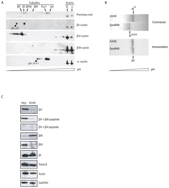
Figure 1. Isotype-specific immunoreactivity of a human βV-tubulin antibody.
A) Taxol-pelleted microtubules from Hey cells were resolved by isoelectric focusing using IPG strips at pH 4.5–5.5, followed by SDS-PAGE, total protein staining and immunoblotting with isotype specific antibodies as indicated. B) Isoelectric focusing of wild type and mutant βI-tubulin in A549 and A549.EpoB40, respectively. IEF gels were either stained with Coomassie blue (top), or electrotransfered onto nitrocellulose for immunoblotting with anti human βV-tubulin antibody (bottom). C) Pre-incubation of antibodies with βIII- or βV-tubulin C-terminal peptides and subsequent immunoblotting using the βV-tubulin antibody on Hey and A549 cell lysates. Also shown are immunoblots with the indicated isotype specific antibodies for βV-, βIII-, βI-, and total β-tubulin.

