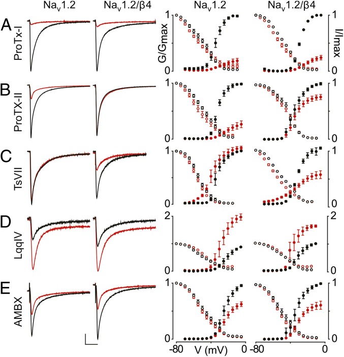
Fig. 1.
Influence of β4 on the ligand susceptibility of Nav1.2. (A–E) Effect of saturating concentrations [100 nM ProTx-I (A), 100 nM ProTx-II (B), 500 nM TsVII (C), 100 nM LqqIV (D), and 500 μM ambroxol (AMBX) (E)] (34, 77) on Nav1.2 and Nav1.2/β4. (Left) Representative sodium currents are elicited by a depolarization to −20 mV before (black) and after (red) addition of toxin or drug from a holding potential of −90 mV. The x-axis is 10 ms; the y-axis is ∼0.5 μA. (Right) Normalized conductance–voltage relationships (G/Gmax; black filled circles) and steady-state inactivation relationships (I/Imax; black open circles) of the WT Nav1.2 channel with or without β4 coexpression are compared before (black circles) and after (red circles) toxin or drug application. β4 alters Nav1.2 susceptibility to ProTx-II and TsVII, whereas ProTx-I, LqqIV, and AMBX are not affected. Channel-expressing oocytes were depolarized in 5-mV steps from a holding potential of −90 mV. Boltzmann fit values are reported in Table S1. n = 3–5; error bars represent S.E.M.

