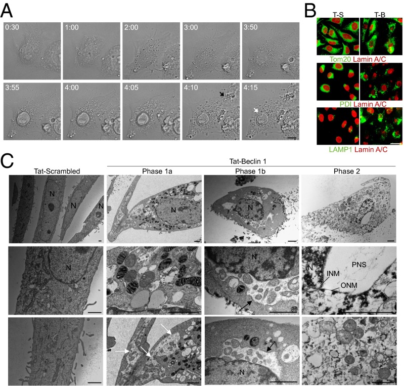
Fig. 3.
Morphological features of Tat-Beclin 1-induced autosis. (A) Representative live-cell imaging of HeLa cells treated with 25 µM Tat-Beclin 1 for 5 h (Movie S1; times shown as hh:mm). The black arrow denotes released intracellular components from a ruptured cell membrane and the white arrow denotes perinuclear space between the inner nuclear membrane and cytoplasm at a region of nuclear concavity. (Scale bar, 10 µm.) (B) Representative images of mitochondrial (Tom20), ER (PDI), late endosome/lysosome (LAMP1), and nuclear lamin-A/C staining in HeLa cells treated with Tat-Scrambled (T-S) or Tat-Beclin 1 (T-B) (20 µM, 5 h). (Scale bar, 20 µm.) (C) EM analysis of HeLa cells treated with peptide (20 µM, 5 h). White arrows show dilated and fragmented ER; black arrows show regions where the perinuclear space has swollen and contains clumps of cytoplasmic material. (Scale bars, 1 µm.) See also Fig. S3.

