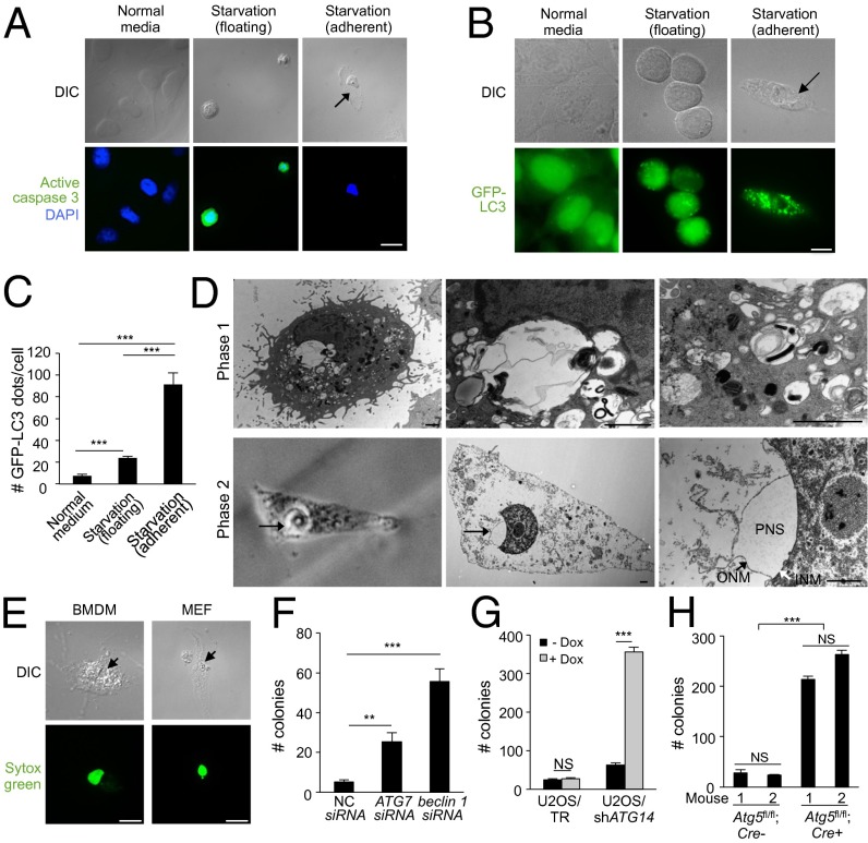
Fig. 4.
Starvation induces autosis. (A) Representative images of active caspase-3 staining in HeLa cells 48 h after starvation (HBSS). (Center) Active caspase 3-positive floating cells with rounded nuclei. (Right) Active caspase 3-negative adherent cell with concave nucleus and swollen perinuclear space. (Scale bar, 20 µm.) (B and C) Representative images (B) and quantitation (C) of GPF-LC3 dots (autophagosomes) in HeLa/GFP-LC3 cells (>50 cells analyzed per sample) grown in normal medium or in floating and adherent HeLa/GFL-LC3 cells 6 h after starvation. (Scale bar, 10 µm.) (D) (Upper) EM images of phase-1 substrate-adherent HeLa cell 6 h after starvation. (Lower) CLEM images of phase-2 substrate-adherent HeLa cell with concave nucleus and swollen perinuclear space (PNS) (arrow) 8 h after starvation. (Lower Left) Phase contrast microscopy; (Lower Center and Lower Right) EM of same cell. The black arrow in Right Lower shows outer nuclear membrane (ONM) and the white arrow shows inner nuclear membrane (INM). (Scale bars, 1 µm.) (E) Representative images of a Sytox Green-positive adherent primary murine BMDM and MEF 48 h after starvation. (Scale bar, 10 µm.) (F) Clonogenic survival of siRNA-transfected adherent HeLa cells starved for 48 h. NC, nontargeting control siRNA. (G) Clonogenic survival of doxycycline (Dox)-inducible adherent U2OS/TR and U2OS/shATG14 cells ± Dox treatment (1 µg/mL) for 5 d before starvation for 72 h. (H) Clonogenic survival of adherent BMDMs (two mice per genotype; Atg5fl/fl;Lyz-Cre− and Atg5fl/fl;Lyz-Cre+ littermates) starved for 72 h. For C and F–H, error bars represent mean ± SEM of triplicate samples and similar results were observed in three independent experiments. For A, B and E, arrows denote concave nucleus and swollen perinuclear space. NS, not significant; **P < 0.01; ***P < 0.001; t test. See also Fig. S4.

