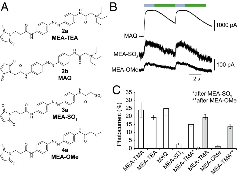
Fig. 4.
Critical role of the photoswitch-positive charge for labeling P2X2 channels. (A) Chemical structures of MEA-TMA analogs. (B) Whole-cell currents evoked by light in cells expressing the I328C mutant tethered with MAQ, MEA-SO3, or MEA-OMe, as indicated. (C) Bar plot showing photocurrent amplitudes elicited by 525-nm light normalized to currents evoked by a saturating concentration of ATP (recorded at 365-nm light) in cells expressing the I328C mutant tethered to the indicated MEA-TMA analogs. Labeling was performed with either 200 μM (white bars) or 80 μM (gray bars) of the analogs (n = 3–7 cells; mean ± SEM).

